Журнал «Травма» Том 18, №4, 2017
Вернуться к номеру
Залишкова фіксованість хребтових сегментів при вибухових переломах грудопоперекового відділу хребта
Авторы: Попсуйшапка К.О., Карпінський М.Ю., Тесленко С.О., Карпінська О.Д., Попов А.І.
ДУ «Інститут патології хребта та суглобів ім. проф. М.І. Ситенка НАМН України», м. Харків, Україна
Рубрики: Травматология и ортопедия
Разделы: Клинические исследования
Версия для печати
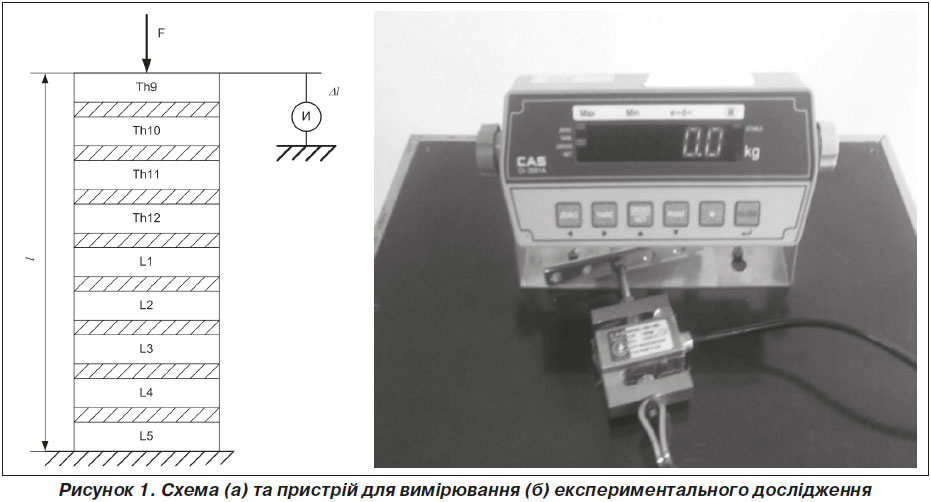
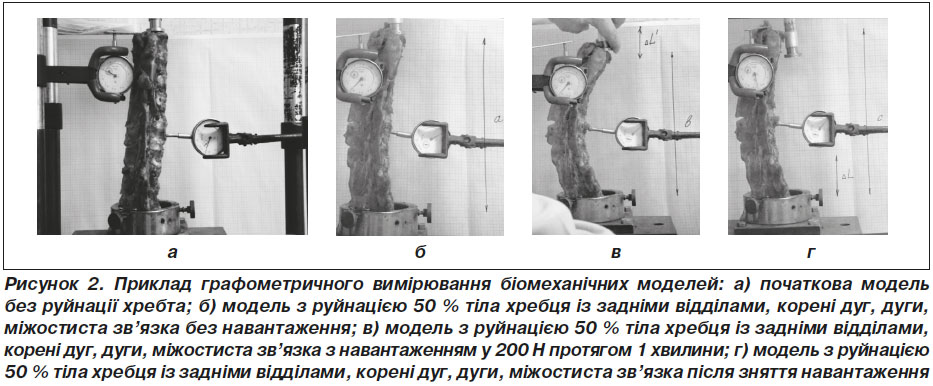
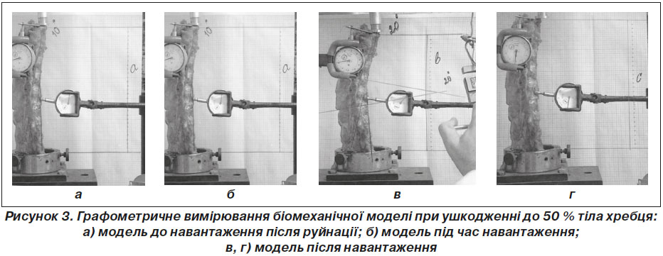
Вступ. Вибухові переломи є найбільш поширеними та найбільш тяжкими ушкодженнями хребта. Вибухові переломи характеризуються багатовідламковим ураженням тіла хребця, можуть супроводжуватись розривом зв’язок і переломом елементів заднього опорного комплексу з формуванням різних видів деформацій, а саме з пружною та пластичною. Мета. Експериментально визначити залишкову фіксованість та залишкову деформацію при вибухових переломах грудопоперекового відділу хребта. Матеріали та методи. Предметом даного дослідження стала фізична модель вибухового перелому тіла Th12 хребця. Було створено шість моделей на анатомічних препаратах блоків хребетних сегментів тварини (свиня) на протязі від ThIX до LV хребців. Блоки хребців забирались у статевозрілих тварин однакового віку. Довжина блоків становила 200 мм. Перша модель була представлена хребетними сегментами зі збереженими кістковими та зв’язковими структурами (I група — норма). У другій моделі було зруйновано до 50 % тіла хребця й один суміжний міжхребцевий диск, при збереженні задніх відділів хребта та коренів дуг (II група — 30–50 % тіл). У третій моделі було зруйновано 50 % тіла хребця, один суміжний міжхребцевий диск, а також задні відділи хребта та корені дуг (III група — 50 % тіла). У четвертій моделі було зруйновано 50 % тіла хребця, один суміжний міжхребцевий диск і задні відділи тіла хребця, корені дуг, дуги, міжостисті зв’язки (IV група). В п’ятій моделі зруйновано все тіло (100 %) і один суміжний диск (V група — 100 % тіла). У шостій групі було зруйновано 100 % тіла хребця, диск, дуги та зв’язки (VI група). Моделі випробували вертикальним осьовим навантаженням величиною 200 Н. Вимірювали величину кіфотичної деформації, осьового подовження після зняття навантаження. Результати. Під час дослідження були отримані дані про зміни величин кіфотичної деформації хребта при осьовому навантаженні залежно від обсягу руйнування при вибуховому переломі тіла Th12 хребця. Також були вивчені залишкові пружні властивості препаратів хребта при наявності зазначених руйнувань. Висновки. Розроблена експериментальна модель на хребті тварин дозволила нам виявити основні закономірності розвитку кіфотичної деформації при вибухових переломах грудопоперекового відділу хребта, а саме: визначити основні біомеханічні показники — залишкову фіксованість і залишкову деформацію хребта. Ушкодження тіла хребця до 50 % призводить до первинного розвитку залишкової деформації. Подальше навантаження хребта не призводить до прогресування деформації, і залишкова фіксованість хребта становить 100 %. Ушкодження заднього опорного комплексу хребта призводить до незначної прогресії залишкової деформації до 6 %.
Введение. Взрывные переломы являются наиболее распространенными и наиболее тяжелыми повреждениями позвоночника. Взрывные переломы характеризуются многооскольчатыми повреждениями тела позвонка, могут сопровождаться разрывом связок и переломом элементов заднего опорного комплекса с формированием разных видов деформации, упругого и пластичного характера. Цель. Экспериментально определить остаточную фиксированность и остаточную деформацию при взрывных переломах грудопоясничного отдела позвоночника. Материалы и методы. Предметом данного исследования была физическая модель взрывного перелома тела Th12 позвонка. Были созданы шесть моделей на анатомических препаратах блоков позвоночных сегментов животных (свинья) на протяжении от ThIX до LV позвонков. Блоки позвонков забирались от половозрелых животных одного возраста. Длина блоков составляла 200 мм. Первая модель была представлена позвоночными сегментами с целыми костными и связочными структурами (I группа — норма). На второй модели были разрушены до 50 % тела позвонка и один межпозвонковый диск, при сохранении задних отделов позвонка и корней дуг (II группа — 30–50 % тела). В третьей модели были разрушены до 50 % тела позвонка, один смежный межпозвонковый диск, задние отделы позвонка и корни дуг (III группа — 50 % тела). В четвертой модели были разрушены 50 % тела позвонка, один смежный межпозвонковый диск, задние отделы позвонка, корни дуг, дуги, меж-остистые связки (IV группа). В пятой модели все тело (100 %) и один смежный диск разрушены (V группа — 100 % тела). В шестой модели разрушены 100 % тела позвонка, диск, дуги и связки (VI группа). Модели испытывали вертикальной осевой нагрузкой величиной 200 Н. Измеряли величину кифотической деформации, осевого удлинения после снятия нагрузки. Результаты. В ходе исследования были получены данные об изменении величины кифотической деформации позвоночника при осевой нагрузке в зависимости от объема разрушений при взрывном переломе тела Th12 позвонка. Также были изучены остаточные упругие свойства препаратов хребта при наличии указанных разрушений. Выводы. Разработанная экспериментальная модель на позвоночнике животного позволила определить основные закономерности развития кифотической деформации при взрывных переломах грудопоясничного отдела позвоночника, а именно: определить основные биомеханические показатели — остаточную фиксированность и остаточную деформацию позвоночника. Повреждение тела позвонка до 50 % приводит к первичному развитию остаточной деформации. Дальнейшая нагрузка на позвоночник не приводит к прогрессированию деформации, и остаточная фиксация составляет 100 %. Повреждение заднего опорного комплекса позвоночника приводит к незначительной прогрессии остаточной деформации до 6 %.
Background. Burst fractures are the most common and most serious injuries of the spine. Burst fractures are characterized by multisplintered injuries of the vertebral body, may be accompanied by rupture of ligaments and by fractures of the elements of the posterior support complex. The purpose of the study was to determine experimentally the residual fixation and permanent deformation in burst fractures of the thoracolumbar spine. Materials and methods. Subject of this study is the physical model of the Th12 vertebral body burst fracture. There were created six models using anatomical preparation of the vertebral segment blocks of the animal (pig) in length from ThIX to LV vertebrae. The vertebral blocks were collected from sexually mature animals of the same age. The length of the blocks was 200 mm. The first model was presented by vertebral segments of saved bone and ligamentous structures (group I — normal). The second model had 50 % of destroyed vertebral body and one adjacent intervertebral disc, keeping the spine and posterior roots of arcs (II group — 30–50 % of the body). There were destroyed 50 % of the vertebral body, one adjacent intervertebral disc, posterior spine section and roots of arcs of the third model (III group — 50 % of the body). There were destroyed 50 % of the vertebral body, one adjacent intervertebral disc, posterior vertebral body, roots of arcs, arcs, interspinous ligaments (IV group). There were destroyed 100 % of the entire body and one adjacent disc of the fifth model (V group — 100 % destruction of the body). In the sixth group, there were destroyed 100 % of the vertebral body, disc, arcs and interspinous ligaments (VI group). Models were tested with vertical axial load value 200 N. We measured the value of kyphotic deformation of axial elongation after removing the load. Results. The initial length of the object with entire bone structure (the rate) was 175 mm. Sagittal balance parameters have negative values — local angular deformation is –5°. The length of the model (II group — 30–50 % of the body) with damage up to 50 % the vertebral body, without damage or with minimal damage to the posterior vertebral body, without damage of the roots of arcs, was 150 mm. Kyphotic deformation — 10°. With load of 200 N, kyphotic deformation value was up to 20°, and the length was 145 mm. When removing the load, the length of the object completely returned to the initial level of the damaged model — to 150 mm. The residual deformation that emerged due to the vertebral body destruction caused the loss in the height by 25 mm of the object without destruction, and its value was 14.3 %. Local kyphotic deformation increased by 15 % from –5 to +10° as compared to the initial state of the object without deformation. With load of 200 N, kyphotic deformation value increased from 10° to 20°, and length of the object was 145 mm. When removing the load, the kyphotic deformation value and the length of the object have completely returned to 150 mm and 10°, respectively. Thus, the residual fixation of the spine at 50 % damage of the vertebral body with preserved posterior vertebral arcs and roots is 100 %, and the residual spine deformation remained 14.3 % from the initial state of the body. This determines the fact that it is the primary spine destruction causes residual deformation, and further load does not lead to the development of deformations, and damaged spine is fully resilient deformation option. 50 % of the vertebral body, one adjacent cranial intervertebral disc, posterior vertebral sections and roots of arcs were destroyed during the study of the next model (III group — 50 % of the body). In comparison with previous investigation, the length of the object has not changed. The load of 200 N caused angular deformation up to 40°, and the length of the object was 135 mm. After unloading, the length returned to 150 mm. It means that residual fixation at 50 % vertebral body deformation with posterior vertebral sections and roots of arcs is 100 %, and residual strain of the spine in comparison with initial undamaged state of the object remained the same — 14.3 %. The next model is characterized by 50 % of vertebral body deformation with posterior vertebral sections and roots of arcs, arcs and interspinous ligament destruction. The length of the object was 150 mm. Under the loading with 200 N, the angular deformation became 42° and the length — 135 mm. After the loading has been removed, the length of the object has changed by 5 mm from the state before loa-ding and was 145 mm. Thus, the damage to the posterior sections of the spine didn’t change the length of the model. After loading, the residual spine fixation became 96.5 %, and residual strain increased to 17.8 % in comparison with the initial state of the object. Next model was characterized by 100 % damage of the vertebral body and one adjacent disc. Initial length of the object in normal state was 200 mm. After destruction of the vertebral body by 100 %, it has been changed to 155 mm. The residual strain of the vertebral body has been changed to 21.5 %. This residual strain progressed due to subsidence of osteal fragments at the destruction of the vertebral body. Then, the load of 200 N caused the deformity with 24° angle. The length of the model under loading was 145 mm. After removing the load, the length of the model returned to the initial level. Thus, the residual fixation of the vertebral body after its 100 % damage (loading power 200 N) is 100 %, and residual strain of the spine was changed to 21.5 % in comparison with the initial state of the object. Group VI was presented by vertebral body destroyed by 100 %, intervertebral ligament rupture, arc fracture. At these injuries, the residual strain of the spine remained 21.5 % in accordance with the initial state of the object. At loading power of 200 N, the deformation angle increased to 44°, and the length of the model reduced from 135 to 155 mm. After unloading, the length of the model became 147 mm. Thus, residual deformity of the vertebral body increased to 26.7 % from the initial state of the object. Conclusions. Experimental model designed on the spine of the animal helped us to determine basic patterns of kyphotic deformity in burst thoracolumbar spine fractures. Particularly, to identify the main biomechanical characteristics: residual fixation and residual deformation of the spine. Destruction of the vertebral body to 50 % leads to primary development of residual deformation. The further load on the vertebral body doesn’t lead to deformity progression, and residual fixation of the vertebral body is 100 %. Damage to the posterior spinal support complex causes a slight progression of the residual deformation
up to 6 %.
хребет; вибуховий перелом; залишкова деформація
позвоночник; взрывной перелом; остаточная деформация
spine; burst fracture; residual deformation


