Інформація призначена тільки для фахівців сфери охорони здоров'я, осіб,
які мають вищу або середню спеціальну медичну освіту.

Підтвердіть, що Ви є фахівцем у сфері охорони здоров'я.

Журнал «Травма» Том 24, №2, 2023

Вернуться к номеру

Математичне моделювання варіантів транспедикулярної фіксації ділянки грудопоперекового переходу після резекції хребця Тh12 під впливом стискаючого навантаження

Авторы: Нехлопочин О.С. (1), Вербов В.В. (1), Чешук Є.В. (1), Карпінський М.Ю. (2), Яресько О.В. (2)
(1) — ДУ «Інститут нейрохірургії ім. акад. А.П. Ромоданова НАМН України», м. Київ, Україна
(2) — ДУ «Інститут патології хребта та суглобів ім. проф. М.І. Ситенка НАМН України», м. Харків, Україна

Рубрики: Травматология и ортопедия

Разделы: Клинические исследования

Версия для печати


Резюме

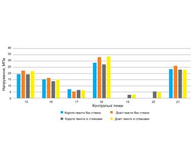
Актуальність. Ділянка грудопоперекового переходу характеризується значним навантаженням, що пред’являє підвищені вимоги до стабілізації, яка повинна не тільки визначати надійну та жорстку фіксацію, але й забезпечувати максимально рівномірний розподіл навантаження на всі елементи як металоконструкції, так і кісткової тканини, з метою виключення неспроможності фіксації в довгостроковій перспективі. Мета: дослідити вплив довжини транспедикулярного гвинта та наявності поперечних стяжок на особливості розподілу навантаження при хірургічній резекції одного хребця зони грудопоперекового переходу під впливом осьового стискаючого навантаження. Матеріали та методи. Проаналізована математична скінченно-елементна модель ділянки грудопоперекового відділу хребта людини (Th9-L5), де хребець Тh12 був видалений та заміщений міжтіловою опорою із додатковою фіксацією транспедикулярною системою. Моделювали 4 варіанти транспедикулярної фіксації з використанням коротких та довгих гвинтів, а також з використанням двох поперечних стяжок та без них. Напружено-деформований стан моделей досліджували під впливом вертикального стискаючого розподіленого навантаження 350 Н. Результати. При використанні коротких гвинтів та за відсутності поперечних стяжок максимальні напруження в хребцях Тh10, Тh11, L1 та L2 становлять відповідно 7,2; 5,3; 4,2 та 14,3 МПа. При використанні довгих гвинтів без стяжок — 6,5; 4,6; 3,8 та 13,5 МПа відповідно. Модель із короткими гвинтами та стяжками демонструє 7,1; 4,4; 3,9 та 14,0 МПа, тоді як застосування довгих гвинтів зі стяжками — 6,3; 4,5; 3,5 та 13,2 МПа відповідно. Висновки. При стискаючому навантаженні використання довгих гвинтів дозволяє знизити рівень напружень у кісткових елементах моделей, використання поперечних стяжок надає більшої жорсткості задньому опорному комплексу транспедикулярної конструкції, що призводить до підвищення напружень на фіксуючих гвинтах, але дозволяє знизити рівень напружень у кістковій тканині.

Background. The area of the thoracolumbar junction is characterized by a significant load that dictates increased requirements to stabilization, which should not only provide a reliable and rigid fixation, but also ensure the maximum uniform distribution of the load on all elements of both the metal structure and the bone tissue to exclude the failure of fixation in the long run. Purpose of the study is to investigate the influence of the transpedicular screw length and the presence of crosslinks on the load distribution during surgical resection of one vertebra from the thoracolumbar junction under the influence of axial compressive load. Materials and methods. We analyzed mathematical finite-element model of the part of thoracolumbar spine (Th9-L5), where the Th12 vertebra was removed and replaced by an interbody implant with additional fixation by a transpedicular system. Four variants of transpedicular fixation were modeled using short and long screws, as well as with and without two crosslinks. The stress-strain state of the models was studied under the influence of a vertical compressive distributed load of 350 N. Results. When using short screws and in the absence of crosslinks, the maximum stresses in the Th10, Th11, L1, and L2 vertebrae are 7.2, 5.3, 4.2, and 14.3 MPa, respectively, when using long screws without crosslinks — 6.5, 4.6, 3.8 and 13.5 MPa. The model with short screws and crosslinks shows 7.1, 4.4, 3.9 and 14.0 MPa, while the application of long screws with crosslinks is 6.3, 4.5, 3.5 and 13.2 MPa, respectively. Conclusions. With a compressive load, the use of long screws allows to reduce the level of stress in the bone elements of the models, the use of crosslinks provides greater rigidity to the posterior support of the transpedicular structure, which leads to an increase in stress on the fixing screws but allows to reduce the level of stress in the bone tissue.


Ключевые слова

скінченно-елементна модель; грудопоперековий перехід; корпоректомія; транспедикулярна стабілізація; поперечна стяжка

finite element model; thoracolumbar junction; corpectomy; transpedicular fixation; crosslink


Для ознакомления с полным содержанием статьи необходимо оформить подписку на журнал.


Список литературы

1. Leucht P., Fischer K., Muhr G., Mueller E.J. Epidemio-logy of traumatic spine fractures. Injury. 2009. 40(2). 166-172. doi: 10.1016/j.injury.2008.06.040, PMID: 19233356.
2. Oliver M., Inaba K., Tang A., Branco B.C., Barmparas G., Schnuriger B., et al. The changing epidemiology of spinal trauma: a 13-year review from a Level I trauma centre. Injury. 2012. 43(8). 1296-1300. doi: 10.1016/j.injury.2012.04.021, PMID: 22648015.
3. Hu R., Mustard C.A., Burns C. Epidemiology of incident spinal fracture in a complete population. Spine (Phila Pa 1976). 1996. 21(4). 492-499. doi: 10.1097/00007632-199602150-00016, PMID: 8658254.
4. Vaccaro A.R., Oner C., Kepler C.K., Dvorak M., Schnake K., Bellabarba C., et al. AOSpine thoracolumbar spine injury classification system: fracture description, neurological status, and key modifiers. Spine (Phila Pa 1976). 2013. 38(23). 2028-2037. doi: 10.1097/BRS.0b013e3182a8a381, PMID: 23970107.
5. Altay M., Ozkurt B., Aktekin C.N., Ozturk A.M., Dogan O., Tabak A.Y. Treatment of unstable thoracolumbar junction burst fractures with short- or long-segment posterior fixation in magerl type a fractures. Eur. Spine J. 2007. 16(8). 1145-1155. doi: 10.1007/s00586-007-0310-5, PMID: 17252216.
6. Müller U., Berlemann U., Sledge J., Schwarzenbach O. Treatment of thoracolumbar burst fractures without neurologic deficit by indirect reduction and posterior instrumentation: bisegmental stabilization with monosegmental fusion. Eur. Spine J. 1999. 8(4). 284-289. doi: 10.1007/s005860050175, PMID: 10483830.
7. Dai L.Y., Jiang S.D., Wang X.Y., Jiang L.S. A review of the management of thoracolumbar burst fractures. Surg. Neurol. 2007. 67(3). 221-231; discussion 231. doi: 10.1016/j.surneu.2006.08.081, PMID: 17320622.
8. Xu H.Z., Wang X.Y., Chi Y.L., Zhu Q.A., Lin Y., Huang Q.S., et al. Biomechanical evaluation of a dynamic pe-dicle screw fixation device. Clin. Biomech. (Bristol, Avon). 2006. 21(4). 330-336. doi: 10.1016/j.clinbiomech.2005.12.004, PMID: 16434133.
9. McNamara M.J., Stephens G.C., Spengler D.M. Transpedicular short-segment fusions for treatment of lumbar burst fractures. J. Spinal Disord. 1992. 5(2). 183-187. doi: 10.1097/00002517-199206000-00006, PMID: 1606376.
10. McLain R.F., Sparling E., Benson D.R. Early failure of short-segment pedicle instrumentation for thoracolumbar fractures. A preliminary report. J. Bone Joint Surg. Am. 1993. 75(2). 162-167. doi: 10.2106/00004623-199302000-00002, PMID: 8423176.
11. Fradet L., Petit Y., Wagnac E., Aubin C.E., Arnoux P.J. Biomechanics of thoracolumbar junction vertebral fractures from various kinematic conditions. Medical & biological engineering & computing. 2014. 52(1). 87-94. doi: 10.1007/s11517-013-1124-8, PMID: 24165806.
12. Verheyden A.P., Spiegl U.J., Ekkerlein H., Gercek E., Hauck S., Josten C., et al. Treatment of Fractures of the Thoracolumbar Spine: Recommendations of the Spine Section of the German Society for Orthopaedics and Trauma (DGOU). Global Spine J. 2018. 8(2 Suppl.). 34S-45S. doi: 10.1177/2192568218771668, PMID: 30210959.
13. Matsuzaki H., Tokuhashi Y., Matsumoto F., Hoshino M., Kiuchi T., Toriyama S. Problems and solutions of pedicle screw plate fixation of lumbar spine. Spine (Phila Pa 1976). 1990. 15(11). 1159-1165. doi: 10.1097/00007632-199011010-00014, PMID: 2267611.
14. Cowin S.C. Bone Mechanics Handbook. 2nd ed. Boca Raton: CRC Press, 2001.
15. Boccaccio A., Pappalettere C. Mechanobiology of Fracture Healing: Basic Principles and Applications in Orthodontics and Orthopaedics. In: Klika V., editor. Theoretical Biomecha-nics. 2011.
16. Nekhlopochin A., Nekhlopochin S., Karpinsky M., Shvets A., Karpinskaya E., Yaresko A. Mathematical Analysis and Optimization of Design Characteristics of Stabilizing Vertebral Body Replacing Systems for Subaxial Cervical Fusion Using the Finite Element Method. Hirurgiâ pozvonočnika. 2017. 14(1). 37-45. doi: 10.14531/ss2017.1.37-45.
17. Radchenko V.A., Kutsenko V.A., Popov A.I., Karpinskуi M.Y., Karpinska O.D. Моделювання варіантів транспедикулярної фіксації грудного відділу хребта при резекції одного-трьох хребців. Trauma. 2022. 18(5). 95-102. doi: 10.22141/1608-1706.5.18.2017.114125.
18. Niinomi M. Mechanical biocompatibilities of titanium alloys for biomedical applications. J. Mech. Behav. Biomed. Mater. 2008. 1(1). 30-42. doi: 10.1016/j.jmbbm.2007.07.001, PMID: 19627769.
19. Образцов И.Ф., Адамович И.С., Барер И.С. Проблемы прочности в биомеханике: учеб. пособие для технич. и биолог. спец. ВУЗ. Москва: Высшая школа, 1988.
20. Зенкевич О.К. Метод конечных элементов в технике. Москва: Мир, 1975. 271 с.
21. Алямовский А.А. SolidWorks/COSMOSWorks. Инженерный анализ методом конечных элементов. Москва: ДМК Пресс, 2004. 432 с.

Вернуться к номеру