
Журнал «Медицина неотложных состояний» Том 20, №8, 2024
Вернуться к номеру
Ультразвукова діагностика вогнепальних ушкоджень великих артеріальних судин шиї
Авторы: Абдуллаєв Р.Я. (1), Лурін І.А. (2, 3), Гречаник О.І. (4), Абдуллаєв Р.Р. (1), Посохов М.Ф. (5), Ібрагімова К.Н. (1), Казмірчук К.А. (4)
(1) - Харківський національний медичний університет, м. Харків, Україна
(2) - Національна академія медичних наук України, м. Київ, Україна
(3) - ДНУ «Центр інноваційних технологій охорони здоров’я» ДУС, м. Київ, Україна
(4) - Національний військово-медичний клінічний центр «Головний військовий клінічний госпіталь», м. Київ, Україна
(5) - ДУ «Інститут неврології, психіатрії та наркології НАМН України», м. Харків, Україна
Рубрики: Медицина неотложных состояний
Разделы: Клинические исследования
Версия для печати
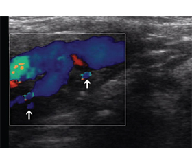
Актуальність. Наявність великих судин, безліч життєво важливих структур та незахищеність шиї робить цю ділянку тіла особливо уразливою для різних травм, зокрема вогнепальних поранень. Мета: оцінити можливості ультрасонографії в діагностиці вогнепальних ушкоджень великих артеріальних судин шиї. Матеріали та методи. Проведено аналіз результатів ультрасонографії у діагностиці вогнепальних ушкоджень великих судин шиї у 53 поранених. Візуалізація великих судин шиї проводилася за допомогою лінійного та мікроконвексного датчиків у частотному діапазоні 5–10 та 4–9 МГц на апараті Philips HD11. Результати. Ушкодження загальної сонної артерії зафіксовано у 38 випадках, внутрішньої сонної артерії — у 23, зовнішньої сонної артерії — у 21, підключичної артерії — у 24, хребетної артерії — у 15. Були виявлені такі типи ушкоджень: відрив інтими з формуванням клаптя — у 27 випадках, відшарування інтими — у 45 випадках, псевдоаневризма —у 49 випадках. Статистично вірогідна відмінність (P < 0,01) була виявлена між кількістю псевдоаневризм та відривом інтими з формуванням клаптя (P < 0,01). Серед ушкоджень загальної сонної артерії псевдоаневризма зустрічалася вірогідно частіше, ніж відрив інтими з формуванням клаптя (P < 0,01) та відшарування інтими (P < 0,05). Пошкодження загальної сонної артерії діагностувалося статистично вірогідно частіше, ніж внутрішньої сонної артерії (P < 0,05) та зовнішньої сонної артерії (P < 0,01), підключичної артерії (P < 0,05), а також хребетної артерії (P < 0,001). Висновки. Ультрасонографія дозволяє виявити ушкодження всіх великих артерій шиї, отримані за бойової травми. Псевдоаневризми статистично вірогідно переважають над відривом інтими з формуванням клаптя та розшаруванням шарів стінки артерій шиї. Ультрасонографія має достатню чутливість (87,8 %), специфічність (60,9 %) і точність (82,6 %) у діагностиці вогнепальних пошкоджень великих артерій шиї, а також у діагностиці псевдоаневризми (93, 75, 90,2 % відповідно).
Background. The presence of large vessels, many vital structures, and the lack of protection make the neck particularly vulnerable to various injuries, including gunshot wounds. The purpose was to evaluate the possibilities of ultrasonography in the diagnosis of gunshot injuries of the major arteries of the neck. Materials and methods. The analysis of the ultrasonography results in diagnosis of gunshot injuries to the large vessels of the neck in 53 injured persons was carried out. Visualization of the large vessels of the neck was carried out using linear and microconvex sensors in the frequency range of 5–10 and 4–9 MHz on the Philips HD11 device. Results. Damage to the common carotid artery was recorded in 38 cases, the internal carotid artery — in 23, the external carotid artery — in 21, the subclavian artery — in 24, the vertebral artery — in 15. The following types of damage were detected: intimal tear with flap formation in 27 cases, detachment of the intima in 45, pseudoaneurysm in 49 cases. A statistically significant difference (P < 0.01) was found between the number of pseudoaneurysms and intimal tear with flap formation (P < 0.01). Among injuries of the common carotid artery, pseudoaneurysm occurred significantly more often than intimal tear with flap formation (P < 0.01) and intimal detachment (P < 0.05). Damage to the common carotid artery was diagnosed statistically significantly more often than to the internal carotid artery (P < 0.05) and external carotid artery (P < 0.01), subclavian artery (P < 0.05), and vertebral artery (P < 0.001). Conclusions. Ultrasonography makes it possible to detect damage to all major arteries of the neck sustained due to combat trauma. Pseudoaneurysms statistically reliably prevail over intimal tear with flap formation and cervical artery dissection. Ultrasonography has sufficient sensitivity (87.8 %), specificity (60.9 %) and accuracy (82.6 %) in the diagnosis of gunshot injuries of the major arteries of the neck, as well as pseudoaneurysm (93, 75, 90.2 %, respectively).
ультрасонографія; великі судини шиї; вогнепальне ушкодження судин шиї
ultrasonography; major arteries of the neck; gunshot damage to vessels of the neck
Для ознакомления с полным содержанием статьи необходимо оформить подписку на журнал.
- Strudwick K, McPhee M, Bell A, Martin-Khan M, Russell T. Review article: Best practice management of neck pain in the emergency department (part 6 of the musculoskeletal injuries rapid review series). Emerg Med Australas. 2018 Dec;30(6):754-772.
- Teixeira F, Menegozzo CA, Netto SD. Safety in selective surgical exploration in penetrating neck trauma. World J Emerg Surg. 2016;11:32.
- Wang D, Zhao Yi, Cha D, Fang P, Liu Y. Penetrating neck trauma with common carotid artery injury caused by a percussive drill а case report. Medicine. 2019;98:22. doi: 10.1097/MD.0000000000015750.
- Coleman KC, Hudnall A, Daniel J, Pillai L, Borgstrom DC, Wilson A, Bardes JM. Penetrating Trauma to the Neck: Utilizing Your Vascular Toolkit. J Trauma Acute Care Surg. 2021 Aug 1;91(2):e51-e54. doi: 10.1097/TA.0000000000003159. PMCID: PMC8369043.
- Pinto A, Russo A, Reginelli A, Iacobellis F, Di Serafino M, Giovine S, Romano L. Gunshot Wounds: Ballistics and Imaging Findings. Seminar in Ultrasound CT MR. 2019 Feb;40(1):25-35.
- Bodanapally UK, Dreizin D., Sliker CW, Boscak AR, Reddy RP. Vascular Injuries to the Neck After Penetrating Trauma: Diagnostic Performance of 40- and 64-MDCT Angiography. AJR Am J Roentgenol. 2015 Oct;205(4):866-872. doi: 10.2214/AJR.14.14161. PMID: 26397338.
- Klein Y, Arieli I, Sagiv S. Cervical spine injuries in civilian victims of explosions: Should cervical collars be used? J Trauma Acute Care Surg. 2016;80(6):985-988.
- Lebowitz C, Matzon JL. Arterial Injury in the Upper Extremity: Evaluation, Strategies and Anticoagulation Management. Hand Clin. 2018 Feb;34(1):85-95.
- Abdullaiev RYa, Grechanyk EI, Kulikova FI, Khvisiuk AN, Cherednichenkov NA and Kogut AV. Ultrasonography in the Diagnosis of Gunshot Injuries of the Neurovascular Bundle of the Extremities EC Neurology 10.11 (2018).
- Mahmoud MZ, Saadi MA, Abuderman A et al. “To-and-fro” waveform in the diagnosis of arterial pseudoaneurysms. World J Radiol. 2015;7(5):89-99. PMID: 4444605.
- Foreman PM, Griessenauer CJ, Kicielinski KP et al. Reliability assessment of the Biffl Scale for blunt traumatic cerebrovascular injury as detected on computer tomography angiography. J Neurosurg. 2017 Jul;127(1):32-35. PMID: 27767400.
- Crilly SM, McElroy E, Ryan J. et al. “Mixed” trauma to the carotid artery in a mixed martial arts injury -A case report and review of the literature. Radiology Case. 2018 May;12(5):1-11. doi: 10.3941/jrcr.v12i5.3234.
- DeSouza IS, Benabbas R, McKee S, Zangbar B, Jain A, Paladino L, Boudourakis L, Sinert R. Accuracy of Physical Examination, Ankle-Brachial Index, and Ultrasonography in the Diagnosis of Arterial Injury in Patients With Penetrating Extremity Trauma: A Systematic Review and Meta-analysis. Acad Emerg Med. 2017 Aug;24(8):994-1017.
- Abdullaiev RYa, Yefimenko AS, Sysun LA, Logvinenko AV, Posokhov NF, Abdullaie RR, Dudnik TA, Kyrychenko AQ, Tomakh NV. Sonographic characteristics of carotid atherosclerosis in patients with ischemic stroke. Azerbaijan Medical Journal. 2024;1:5-12.
- Stefanopoulos PK, Pinialidis DE, Hadjigeorgiou GF, Filippakis KN. Wound ballistics 101: the mechanisms of soft tissue wounding by bullets. Eur J Trauma Emerg Surg. 2017 Oct;43(5):579-586.
- Prichayudh S, Choadrachata-anun J, Sriussadaporn S. Selective management of penetrating neck injuries using “no zone” approach. Injury 2015;49:1720-5.
- Sandstrom CK, Nunez DB. Head and Neck Injuries: Special Considerations in the Elderly Patient. Neuroimaging Clin N Am. 2018 Aug;28(3):471-481.
- Richard SA, Zhang CW, Wu C, Ting W, Xiaodong X. Traumatic Penetrating Neck Injury with Right Common Carotid Artery Dissection and Stenosis Effectively Managed with Stenting: A Case Report and Review of the Literature. Case Rep Vasc Med. 2018;2018:4602743.
- Abdullayev RY, Lurin İA, Belıy VYa, Kulikova Fİ, Zarutski YaL, Solodyanikova Oİ. Ultrasonographic and X-ray diagnosis of abdominal injuries from gunshot wounds. Azerbaijan Medical Journal. 2023;2:21-27.
- Speelman ES, Brocx B, Wilbers JE. The influence of arm positions on abdominal image quality of whole-body computed tomography in trauma: systematic review. Emerg. Radiol. 2020;27:141-150.