Інформація призначена тільки для фахівців сфери охорони здоров'я, осіб,
які мають вищу або середню спеціальну медичну освіту.

Підтвердіть, що Ви є фахівцем у сфері охорони здоров'я.


UkraineNeuroGlobal


UkraineNeuroGlobal

Международный неврологический журнал Том 18, №8, 2022

Вернуться к номеру

Гліобластома: огляд літератури

Авторы: Dipak Chaulagain, V. Smolanka, A. Smolanka, T. Havryliv
Uzhhorod Regional Clinical Center of Neurosurgery and Neurology, Uzhhorod National University, Uzhhorod, Ukraine

Рубрики: Неврология

Разделы: Справочник специалиста

Версия для печати


Резюме

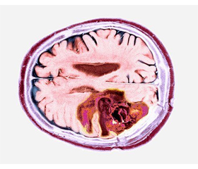
Гліобластома є найбільш агресивним типом пухлини головного мозку, визначена як астроцитома 4-го типу відповідно до 5-го видання класифікації пухлин центральної нервової системи Всесвітньої організації охорони здоров’я. Це рак, що починається в гліальних клітинах, які є підтримуючими клітинами мозку. Гліобластома є рідкісним типом пухлини головного мозку і становить близько 15 % усіх цих пухлин. Найчастіше вона зустрічається в дорослих віком понад 50 років, причому в чоловіків розвивається частіше, ніж у жінок. Симптоми гліобластоми можуть відрізнятися залежно від локалізації пухлини в головному мозку. Загальні симптоми включають головний біль, нудоту, блювання та зміни зору. Іншими симптомами можуть бути судоми, зміни в поведінці або особистості, а також труднощі з мовою або рухами. Стандартом лікування гліобластоми є поєднання хірургічного втручання, променевої та хіміотерапії. Хірургічне втручання є першим кроком у лікуванні, метою якого є видалення якомога більшої частини пухлини. Променева та хіміотерапія зазвичай використовуються для знищення будь-яких залишкових ракових клітин. Однак, незважаючи на ці методи лікування, прогноз при гліобластомі, як правило, поганий, більшість пацієнтів живуть менше двох років після встановлення діагнозу. Загалом гліобластома є дуже агресивною пухлиною головного мозку з поганим прогнозом. Стандартне лікування передбачає поєднання хірургічного втручання, променевої та хіміотерапії. Останні дослідження були зосереджені на розробці нових методів лікування, таких як цільова терапія та імунотерапія, що показали обнадійливі результати в клінічних випробуваннях.

Glioblastoma is the most aggressive type of brain tumor classified as type 4 astrocytoma according to 5th edition of the World Health Organization сlassification of tumors of the central nervous system. It is a cancer that begins in the glial cells, which are the supporting cells of the brain. Glioblastoma is a rare type of brain tumor and accounts for about 15 % of all brain tumors. It is most commonly found in adults over the age of 50, and men are more likely to develop it than women. The symptoms of glioblastoma can vary depending on the location of the tumor in the brain. Common symptoms include headaches, nausea, vomiting, and changes in vision. Other symptoms can include seizures, changes in behavior or personality, and difficulty with speech or movement. The standard treatment for glioblastoma is a combination of surgery, radiation therapy, and chemotherapy. Surgery is usually the first step in treatment, with the goal of removing as much of the tumor as possible. Radiation therapy and chemotherapy are typically used to kill any remaining cancer cells. However, despite these treatments, the prognosis for glioblastoma is generally poor, with most patients surviving for less than two years after diagnosis. Overall, glioblastoma is a highly aggressive brain tumor with poor prognosis. The standard treatment is a combination of surgery, radiation therapy and chemotherapy. Recent research has focused on developing new treatments, such as targeted therapies and immunotherapies, which have shown promising results in clinical trials.


Ключевые слова

гліобластома; гліома; астроцитома

glioblastoma; glioma; astrocytoma


Для ознакомления с полным содержанием статьи необходимо оформить подписку на журнал.


Список литературы

1. Ohgaki H., Kleihues P. Genetic pathways to primary and se-condary glioblastoma. American Journal of Pathology. 2007. 170(5). 1445-1453. doi: 10.2353/ajpath.2007.070011.
2. Kleihues P., Ohgaki H. Primary and secondary glioblastomas: from concept to clinical diagnosis. Neuro-Oncology. 1999. 1(1). 44-51. doi: 10.1093/neuonc/1.1.44.
3. Mansouri A., Karamchandani J., Das S. Molecular Genetics of Secondary Glioblastoma. 2017. doi: 10.15586/codon.glioblastoma.2017.ch2.
4. Zhang X., Zhang W., Cao W.D., Cheng G., Zhang Y.Q. Glioblastoma multiforme: molecular characterization and current treatment strategy (review). Experimental and Therapeutic Medicine. 2012. 3(1). 9-14. doi: 10.3892/etm.2011.367.
5. Nelson Sarah J., Cha Soonmee. Imaging Glioblastoma Multiforme. Cancer Journal. 2003. 9(2). 134-145. doi: 10.1097/00130404-200303000-00009.
6. Altman David A., Atkinson Denis S., Brat Daniel J. Glioblastoma Multiforme. Radiographics. 2007. 27(3). 883-888. doi: 10.1148/rg.273065138.
7. Brown T.J., Brennan M.C., Li M., Church E.W., Brandmeir N.J. et al. Association of the Extent of Resection With Survival in Glioblastoma: A Systematic Review and Meta-analysis. JAMA Oncology. 2016. 2(11). 1460-1469. doi: 10.1001/jamaoncol.2016.1373.
8. Senft C., Franz K., Ulrich C.T., Bink A., Szelényi A., Gasser T. et al. Low field intraoperative MRI-guided surgery of gliomas: a single center experience. Clinical Neurology and Neurosurgery. 2010. 112. 237-43.
9. Li F.Y., Chen X.L., Sai X.Y., Zhang J.S., Hu S., Li J.J. et al. Application of intraoperative magnetic resonance imaging and multimodal navigation in surgical resection of glioblastoma. Chinese Journal of Surgery. 2013. 51. 542-6 (in Chinese).
10. Gandhi S., Tayebi Meybodi A., Belykh E., Cavallo C., Zhao X. et al. Survival Outcomes Among Patients With High-Grade Glioma Treated With 5-Aminolevulinic Acid-Guided Surgery: A Systematic Review and Meta-Analysis. Front. Oncol. 2019. 9. 620. doi: 10.3389/fonc.2019.00620.
11. Stummer W., Stocker S., Novotny A. et al. In vitro and in vivo porphyrin accumulation by C6 glioma cells after exposure to 5-aminolevulinic acid. Journal of Photochemistry and Photobiology B: Biology. 1998 Sep. 45(2–3). 160-169.
12. Ennis S.R., Novotny A., Xiang J. et al. Transport of 5-aminolevulinic acid between blood and brain. Brain Research. 2003 Jan 10. 959(2). 226-234.
13. Mann J., Ramakrishna R., Magge R., Wernicke A.G. Advan-ces in Radiotherapy for Glioblastoma. Frontiers in Neurology. 2018. 8. 748. doi: 10.3389/fneur.2017.00748.
14. Minniti G., Muni R., Lanzetta G., Marchetti P., Maurizi Enrici R. Chemotherapy for Glioblastoma: Current Treatment and Future Perspectives for Cytotoxic and Targeted Agents. Anticancer Research. 2009. 29. 5171-5184.
15. Huang B., Yu Z., Liang R. Effect of long-term adjuvant temozolomide chemotherapy on primary glioblastoma patient survival. BMC Neurol. 2021. 21. 424. doi: 10.1186/s12883-021-02461-9.
16. Wang Zhuo, Yang Guozi, Zhang Yu-Yu, Yao Yan, Dong Li-Hua. A comparison between oral chemotherapy combined with radiotherapy and radiotherapy for newly diagnosed glioblastoma. Medicine. 2017 Nov. 96(44). e8444. doi: 10.1097/MD.0000000000008444.
17. Zhang L., Wu X., Xu T. et al. Chemotherapy plus radiotherapy versus radiotherapy alone in patients with anaplastic glioma: a systematic review and meta-analysis. J. Cancer Res. Clin. Oncol. 2013. 139. 719-26.
18. Baid Ujjwal, Rane Swapnil U., Talbar Sanjay, Gupta Sudeep, Thakur Meenakshi H. et al. Overall Survival Prediction in Glioblastoma With Radiomic Features Using Machine Learning. Frontiers in Computational Neuroscience. 2020. 14. 61. doi: 10.3389/fncom.2020.00061.
19. Witthayanuwat S., Pesee M., Supaadirek C., Supakalin N., Thamronganantasakul K., Krusun S. Survival Analysis of Glioblastoma Multiforme. APJCP. 2018. 19(9). 2613-2617. doi: 10.22034/APJCP.2018.19.9.2613.
20. Yersal Özlem. Clinical outcome of patients with glioblastoma multiforme: single center experience. Journal of Oncological Sciences. 2017. 3(3). 123-126. doi: 10.1016/j.jons.2017.10.005.

Вернуться к номеру