
Архив офтальмологии Украины Том 11, №2, 2023
Вернуться к номеру
Експресія CD34 у тканинах сітківки та вплив блокади тирозинових протеїнкіназ при діабетичній ретинопатії
Авторы: Водяник В.В.
Національний медичний університет імені О.О. Богомольця, м. Київ, Україна
Рубрики: Офтальмология
Разделы: Клинические исследования
Версия для печати
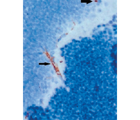
Актуальність. Ангіогенез у сітківці відіграє ключову роль у розвитку та прогресуванні діабетичної ретинопатії (ДР). Ендотеліальні клітини-попередники, що беруть участь у проліферації судин, ідентифікують за допомогою антитіл до CD34, який є маркером ангіогенезу за умов ДР. Мета дослідження: вивчити експресію CD34 у тканинах сітківки та вплив на неї блокади тирозинових протеїнкіназ при розвитку експериментальної діабетичної ретинопатії. Матеріали та методи. У 45 тримісячних щурів-самців лінії Wistar моделювали цукровий діабет шляхом одноразового введення стрептозотоцину в дозі 50 мг/кг (Sigma-Aldrich, Китай). Щурів було розподілено на 3 групи: контрольна, із введенням простого інсуліну і з комбінованим введенням інсуліну й іматинібу в дозі 20 мг/кг у вигляді саше per os (Grindeks, Латвія). Імуногістохімічно у сітківці виявляли CD34 (Thermo Fisher Scietific, США). Результати. Розвиток ДР проявлявся вираженими дегенеративними змінами нервових клітин, що відбувалися на тлі порушень мікроциркуляції у вигляді ішемії, набряку й інтраретинальних судинних аномалій. CD34-позитивні клітини виявлялися: через 7 діб у судинах хоріоїдального сплетення, через 14 діб у розширених судинах шару гангліонарних клітин і через 21 добу у зовнішньому плексіформному шарі. Інтенсивність їх забарвлення збільшувалася. Через 28 діб у контрольній групі поряд з активним ангіогенезом у судинах шару гангліонарних клітин та хоріоїдального сплетення відмічено утворення фіброваскулярних проліфератів, які поширювалися на внутрішній і зовнішній ядерні шари з тенденцією до радіального проростання у навколишні шари сітківки. Введення інсуліну та, більшою мірою, комбінації інсуліну з іматинібом гальмувало розвиток ДР, знижувало інтенсивність CD34-позитивного забарвлення у судинах сітківки та запобігало утворенню фіброваскулярних проліфератів. Висновки. Таким чином, проведене дослідження встановило особливості ангіогенезу й утворення фіброваскулярних проліфератів у сітківці за умов експериментального стрептозотоцинового діабету у щурів. Показаний позитивний вплив блокади тирозинових протеїнкіназ іматинібом щодо встановлених патологічних процесів.
Background. Retinal angiogenesis plays a key role in the development and progression of diabetic retinopathy (DR). Endothelial progenitor cells participating in the vascular proliferation are identified using antibodies to CD34, which is a marker of angiogenesis in DR. The purpose was to study the CD34 retinal expression and the effect on it of tyrosine protein kinase blockade in experimental DR. Material and methods. In 45 three-month-old male Wistar rats, diabetes was simulated by a single injection of streptozotocin (50 mg/kg; Sigma-Aldrich, China). Rats were divided into 3 groups: controls, with the introduction of simple insulin and with the combined administration of insulin and imatinib at a dose of 20 mg/kg in the form of a sachet per os (Grindex, Latvia). CD34 in the retina was detected immunohistochemically (Thermo Fisher Scientific, USA). Results. The development of DR manifested itself by pronounced degenerative changes in nerve cells that occurred against the background of microcirculatory disorders in the form of ischemia, edema, and intraretinal microvascular abnormalities. CD34-positive cells were detected after 7 days in the vessels of the choroid plexus, after 14 days — in the dilated vessels of the ganglion cells layer, and after 21 days — in the outer plexiform layer. The intensity of their staining increased. After 28 days, in the control group, along with active angiogenesis in the vessels of the ganglion cells layer and the choroid plexus, the formation of fibrovascular proliferates was noted, which spread to the inner and outer nuclear layers with a tendency to radial sprouting into the surrounding retinal layers. The introduction of insulin and, to a greater extent, the combination of insulin with imatinib inhibited the development of DR, reduced the intensity of CD34-positive staining in retinal vessels, and prevented the formation of fibrovascular proliferates. Conclusions. Thus, the conducted study revealed the features of angiogenesis and the formation of fibrovascular proliferates in the retina under the conditions of experimental streptozotocin-induced diabetes in rats. The positive effect of tyrosine protein kinase blockade with imatinib on detected pathological processes was shown.
цукровий діабет; стрептозотоцин; ангіогенез; фіброваскулярні проліферати; імуногістохімія; іматиніб
diabetes; streptozotocin; angiogenesis; fibrovascular proliferations; immunohistochemistry; imatinib
Для ознакомления с полным содержанием статьи необходимо оформить подписку на журнал.
- Ogurtsova K., da Rocha Fernandes J.D., Huang Y., Linnenkamp U., Guariguata L., Cho N.H., Cavan D., Shaw J.E., Makaroff L.E. IDF Diabetes Atlas: Global estimates for the prevalence of diabetes for 2015 and 2040. Diabetes Res. Clin. Pract. 2017 Jun. 128. 40-50. doi: 10.1016/j.diabres.2017.03.024.
- Wong T.Y., Sabanayagam C. Strategies to Tackle the Glo–bal Burden of Diabetic Retinopathy: From Epidemiology to Artificial Intelligence. Ophthalmologica. 2020. 243(1). 9-20. doi: 10.1159/000502387.
- Thomas R.L., Halim S., Gurudas S., Sivaprasad S., –Owens D.R. IDF Diabetes Atlas: A review of studies utilising retinal photography on the global prevalence of diabetes related retinopathy between 2015 and 2018. Diabetes Res. Clin. Pract. 2019 Nov. 157. 107840. doi: 10.1016/j.diabres.2019.107840.
- Antonetti D.A., Silva P.S., Stitt A.W. Current understanding of the molecular and cellular pathology of diabetic retinopathy. Nat. Rev. Endocrinol. 2021 Apr. 17(4). 195-206. doi: 10.1038/s41574-020-00451-4.
- Mykheitseva I.M., Molchaniuk N.I., Abdulhadi Muhammad, Kolomiichuk S.G., Suprun O.O. Ultrastructural changes in the chorioretinal complex of the rat after inducing form-deprivation axial myopia only, diabetic retinopathy only and diabetic retinopathy in the presence of myopia. J. Оphthalmol (Ukraine). 2021. 4. 72-78. http://doi.org/10.31288/oftalmolzh202147278.
- Wang W., Lo A.C.Y. Diabetic Retinopathy: Pathophysiology and Treatments. Int. J. Mol. Sci. 2018 Jun 20. 19(6). 1816. doi: 10.3390/ijms19061816.
- Capitão M., Soares R. Angiogenesis and Inflammation Crosstalk in Diabetic Retinopathy. J. Cell. Biochem. 2016 Nov. 117(11). 2443-53. doi: 10.1002/jcb.25575.
- Sidney L.E., Branch M.J., Dunphy S.E., Dua H.S., Hopkinson A. Concise review: evidence for CD34 as a common marker for diverse progenitors. Stem. Cells. 2014 Jun. 32(6). 1380-9. doi: 10.1002/stem.1661.
- Hassanpour M., Salybekov A.A., Kobayashi S., Asahara T. CD34 positive cells as endothelial progenitor cells in biology and medicine. Front. Cell Dev. Biol. 2023 Apr 17. 11. 1128134. doi: 10.3389/fcell.2023.1128134.
- Proia A.D., Caldwell M.C. Intraretinal neovascularization in diabetic retinopathy. Arch. Ophthalmol. 2010 Jan. 128(1). 142-4. doi: 10.1001/archophthalmol.2009.338.
- Zhu J., Sun H., Kang X., Zhu H., Yan X. Acidic polysaccharides from Buddleja officinalis inhibit angiogenesis via the Nrf2/ARE pathway to attenuate diabetic retinopathy. Food Funct. 2022 Aug 30. 13(17). 9021-9031. doi: 10.1039/d2fo01075e.
- Rigato M., Bittante C., Albiero M., Avogaro A., Fadini G.P. Circulating Progenitor Cell Count Predicts Microvascular Outcomes in Type 2 Diabetic Patients. J. Clin. Endocrinol. Metab. 2015 Jul. 100(7). 2666-72. doi: 10.1210/jc.2015-1687.
- Ma J., Zhu T., Tang X., Ye P., Zhang Z. Effect of an intravi–treal injection of bevacizumab on the expression of VEGF and CD34 in the retina of diabetic rats. Clin. Exp. Ophthalmol. 2010 Dec. 38(9). 875-84. doi: 10.1111/j.1442-9071.2010.02370.x.
- Gavi S., Shumay Е., Wang Н., Malbon С. G-protein-coupled receptors and tyrosine kinases: crossroads in cell signaling and regulation. Trends Endocrinol. Metab. 2006. 17(2). 46-52.
- Siddle K. Molecular basis of signaling specificity of insulin and IGF receptors: neglected corners and recent advances. Front Endocrinol (Lausanne). 2012 Feb 28. 3. 34. doi: 10.3389/fendo.2012.00034. PMID: 22649417; PMCID: PMC3355962.
- Pearson G., Robinson F., Beers Gibson T., Xu B.E., Karandikar M., Berman K., Cobb M.H. Mitogen-activated protein (MAP) kinase pathways: regulation and physiological functions. Endocrine Reviews. 2001. 22(2). 153-183. doi: 10.1210/er.22.2.153.
- Liu Y., Chen J., Liang H., Cai Y., Li X., Yan L., Zhou L., Shan L., Wang H. Human umbilical cord-derived mesenchymal stem cells not only ameliorate blood glucose but also protect vascular endothelium from diabetic damage through a paracrine mechanism mediated by MAPK/ERK signaling. Stem Cell Res. Ther. 2022 Jun 17. 13(1). 258. doi: 10.1186/s13287-022-02927-8. PMID: 35715841; PMCID: PMC9205155.
- Hymowitz S.G., Malek S. Targeting the MAPK Pathway in RAS Mutant Cancers. Cold Spring Harb Perspect Med. 2018 Nov 1. 8(11). a031492. doi: 10.1101/cshperspect.a031492.
- Waller C.F. Imatinib Mesylate. Recent Results Cancer Res. 2018. 212. 1-27. doi: 10.1007/978-3-319-91439-8_1.
- Liu Y., Wu N. Progress of Nanotechnology in Diabetic Reti–nopathy Treatment. Int. J. Nanomedicine. 2021 Feb 24. 16. 1391-1403. doi: 10.2147/IJN.S294807.
- Stewart E.A., Samaranayake G.J., Browning A.C., Hopkinson A., Amoaku W.M. Comparison of choroidal and retinal endothelial cells: characteristics and response to VEGF isoforms and anti-VEGF treatments. Exp. Eye Res. 2011 Nov. 93(5). 761-6. doi: 10.1016/j.exer.2011.09.010.
- Wu D., Kanda A., Liu Y., Noda K., Murata M., Ishida S. Involvement of Müller Glial Autoinduction of TGF-β in Diabetic Fibrovascular Proliferation Via Glial-Mesenchymal Transition. Invest. Ophthalmol. Vis. Sci. 2020 Dec 1. 61(14). 29. doi: 10.1167/iovs.61.14.29.
- Fadini G.P., Sartore S., Baesso I., Lenzi M., Agostini C., Tiengo A., Avogaro A. Endothelial progenitor cells and the diabetic paradox. Diabetes Care. 2006 Mar. 29(3). 714-6. doi: 10.2337/dia–care.29.03.06.dc05-1834.
- Liu X., Li Y., Liu Y., Luo Y., Wang D., Annex B.H., Goldschmidt-Clermont P.J. Endothelial progenitor cells (EPCs) mobilized and activated by neurotrophic factors may contribute to pathologic neovascularization in diabetic retinopathy. Am. J. Pathol. 2010 Jan. 176(1). 504-15. doi: 10.2353/ajpath.2010.081152.
- Ding X., Gu R., Zhang M., Ren H., Shu Q., Xu G., Wu H. Microglia enhanced the angiogenesis, migration and proliferation of co-cultured RMECs. BMC Ophthalmol. 2018 Sep 17. 18(1). 249. doi: 10.1186/s12886-018-0886-z.
- Hsu Y.R., Yang C.M., Yeh P.T. Clinical and histological features of epiretinal membrane after diabetic vitrectomy. Graefes Arch. Clin. Exp. Ophthalmol. 2014 Mar. 252(3). 401-10. doi: 10.1007/s00417-013-2479-0.
- Pu X., Zhu P., Zhou X., He Y., Wu H., Du L., Gong H., Sun X., Chen T., Zhu J., Xu Q., Zhang H. CD34+ cell atlas of main organs implicates its impact on fibrosis. Cell. Mol. Life Sci. 2022 Oct 31. 79(11). 576. doi: 10.1007/s00018-022-04606-6.