Международный неврологический журнал Том 20, №5, 2024
Вернуться к номеру
Моделювання тракційної травми периферичного нерва в експерименті
Авторы: Вороді М.В. (1, 2), Петрів Т.І. (1), Нехлопочин О.С. (1), Цимбалюк В.І. (3)
(1) - ДУ «Інститут нейрохірургії імені академіка А.П. Ромоданова НАМН України», м. Київ, Україна
(2) - Національний медичний університет імені О.О. Богомольця, м. Київ, Україна
(3) - Національна академія медичних наук України, м. Київ, Україна
Рубрики: Неврология
Разделы: Клинические исследования
Версия для печати
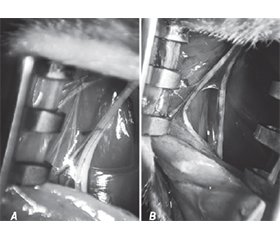
Актуальність. Ушкодження периферичного нерва можуть призвести до значних функціональних порушень і зниження якості життя. Функціональне відновлення периферичного нерва є складним процесом, що залежить від багатьох факторів, деякі з яких можна контролювати для поліпшення результатів одужання. Кількість поранених із ушкодженням периферичних нервів кінцівок може становити до 25 % в умовах війни. Ступінь інвалідизації пацієнтів становить 65–70 %, тому питання відновлення периферичних нервів є надзвичайно актуальним, особливо під час війни. Мета: розробити модель тракційного ушкодження периферичного нерва в експерименті, що виникає внаслідок поздовжнього розтягування сідничного нерва із використанням інструмента, модифікованого на основі стандартного ранорозширювача з кремальєрною системою, який забезпечує можливість відтворення травматичних умов, що найточніше імітують реальні клінічні випадки. Матеріали та методи. Дослідження проводили на 20 безпородних щурах-самцях (середня маса 225 ± 55 г), утримуваних у стандартних умовах віварію ДУ «Інститут нейрохірургії ім. акад. А.П. Ромоданова» НАМН України з дотриманням чинних норм біоетики. Тварини були розділені на дві групи: І група тварин (n = 10) — тракційна травма нерва (ТТН), виведення з експерименту через 15 днів з подальшим дослідженням травмованої ділянки нерва за допомогою світлової і електронної мікроскопії. ІІ група тварин (n = 10) — ТТН, виведення з експерименту через 30 днів з подальшим дослідженням травмованої ділянки нерва за допомогою світлової і електронної мікроскопії. Перед операцією, на 15-й та 30-й день після моделювання травми (перед забором нерва для морфологічного аналізу) визначали функціональний індекс сідничного нерва (sciatic functional index, SFI) з використанням тесту «ходьба по доріжці» (walking track test) у І та ІІ групах тварин. Щурів утримували за стандартним 12-годинним циклом світло/темрява і з наданням вільного доступу до їжі та води. Експеримент завершувався виведенням тварин з експерименту шляхом ін’єкції летальної дози тіопенталу натрію. Для більш детального аналізу на 15-й та 30-й день після початку експерименту проводили повторні оперативні втручання для забору відповідних ділянок нерва для морфологічного дослідження. Для точної ідентифікації проксимального кінця нерва використовували методику накладання лігатури на епіневрій, що дозволяло чітко відмежувати досліджувані ділянки для подальшого аналізу. Структурні зміни в нервовій тканині аналізували за допомогою світлової та електронної мікроскопії, що забезпечувало високу деталізацію морфологічних характеристик. Результати. На початку експерименту, до нанесення тракційного ушкодження, SFI у першій групі дорівнював 2,26 ± 0,27, а у другій групі — 3,14 ± 0,53. Через 15 днів після моделювання травми SFI у першій групі дорівнював 68,9 ± 1,61, а у другій групі — 70,31 ± 1,75, різниця між показниками була статистично невірогідною (р ≥ 0,05). При порівнянні SFI до та після травми різниця виявилася статистично вірогідною (р < 0,0014 і р < 0,0032 відповідно). Через 30 днів після моделювання травми SFI у другій групі дорівнював 32,27 ± 1,13 і порівняно із показником на 15-й день після травми статистично значуще відрізнявся на користь показника через 30 днів (р < 0,0026). З точки зору біомеханіки можна виділити три основні типи ушкоджень, коли основним травмуючим механізмом є тракція: еластичне розтягнення, нееластичне (пластичне) розтягнення та розрив. Еластичне розтягнення характеризується поверненням нерва до його початкової довжини після усунення тракції. При подальшому збільшенні сили розтягнення нерв переходить у фазу пластичної деформації, під час якої відбуваються необоротні зміни в його структурі, як-от розрив аксонів, ендоневральної та периневральної оболонки. При перевищенні певного порогу розтягнення (зокрема, коли гачок зміщувався по кремальєрі на позиції 9, 10, 11-го зубців) відбувався розрив нерва. Результати морфологічних досліджень планується детально описати в майбутніх наукових публікаціях. Висновки. 1. Модель тракційної травми периферичного нерва є об’єктивною, про що свідчать результати функціонального індексу сідничного нерва. Ця методика є легковідтворюваною, не потребує дорогого та складного обладнання. 2. Використання моделі тракційної травми периферичного нерва в експерименті з метою вивчення лікувальних впливів при цьому типі ушкодження дозволить розширити розуміння його пато- та морфогенезу та удосконалити лікувальну тактику. 3. Екстраполяція моделі на клінічну практику дозволить поліпшити лікування потерпілих із наслідками бойових ушкоджень периферичних нервів, у яких часто має місце подібний механізм травми.
Background. Peripheral nerve (PN) injuries can lead to significant functional impairment and reduced quality of life. Functional recovery of PN is a complex process dependent on many factors, some of which can be controlled to improve outcomes. The number of wounded people with damage to the peripheral nerves of the limbs can be up to 25 % in war conditions. The degree of disability of patients is 65–70 %, so the issue of restoring peripheral nerves is extremely relevant, especially during wartime. The purpose was to develop a model of PN traction injury in an experiment resulting from longitudinal stretching of the sciatic nerve using a tool modified based on a standard self-retaining retractor, which provides the possibility of reproducing traumatic conditions that most accurately imitate real clinical cases. Materials and methods. The study was conducted on 20 outbreed male rats (average weight of 225 ± 55 g) kept in standard conditions of the vivarium of the State Institution “Romodanov Neurosurgery Institute of the National Academy of Medical Sciences of Ukraine” in compliance with current norms of bioethics. The animals were divided into two groups: the first one (n = 10) — traction injury of the nerve, withdrawal from the experiment after 15 days with further examination of the damaged area with the light and electron microscopy. The second group of animals (n = 10) — traction injury of the nerve, withdrawal from the experiment after 30 days with further examination of the damaged part of the nerve with the help of light and electron microscopy. Before surgery, on the 15th and 30th day after simulating injury (before the nerve harvesting for morphological analysis), the sciatic functional index (SFI) was determined using the walking track test in groups I and II of animals, respectively. Rats were maintained on a standard 12-h light/dark cycle with free access to food and water. The experiment ended by removing the animals from the experiment by injecting a lethal dose of thiopental sodium. For a more detailed analysis, on the 15th and 30th day after the start of the experiment, repeated surgical interventions were performed to collect the appropriate sections of the nerve for morphological examination. For accurate identification of the proximal end of the nerve, the method of applying a ligature to the epineurium was used, which allowed to clearly demarcate the studied areas for further analysis. Structural changes in the nerve tissue were analyzed using light and electron microscopy, which provided high detail of morphological characteristics. Results. At the beginning of the experiment, before traction injury, the SFI in the first group was 2.26 ± 0.27, and in the second group — 3.14 ± 0.53. Fifteen days after injury simulation, the SFI in the first group was 68.90 ± ± 1.61, and in the second group — 70.31 ± 1.75, the difference between the indicators was statistically insignificant (p ≥ 0.05). When comparing the SFI before and after injury, the difference was statistically significant (p < 0.0014 and p < 0.0032, respectively). Thirty days after injury simulation, the SFI in the second group was 32.27 ± 1.13 and, compared to the indicator on the 15th day after the injury, it differed statistically significantly in favor of the indicator after 30 days (p < 0.0026). From the biomechanical point of view, three main types of injuries can be distinguished, when traction is the main traumatic mechanism: elastic stretching, inelastic (plastic) stretching and rupture. Elastic stretching is characterized by the return of the nerve to its original length after removal of traction. With a further increase in the stretching force, the nerve enters the phase of plastic deformation, during which irreversible changes in its structure occur, such as the rupture of axons, endoneural and perineural sheaths. When a certain threshold of stretching is exceeded (in particular, when the hook was moved along racks at position of 9, 10, 11 teeth), nerve rupture occurred. The results of morphological studies are planned to be described in detail in future scientific publications. Conclusions. 1. The model of the peripheral nerve traction injury is objective as evidenced by the sciatic nerve functional index. This technique is easily reproducible and does not require expensive and complex equipment. 2. The use of a model of peripheral nerve traction injury in an experiment, with the aim of studying the therapeutic effects in this type of trauma, will allow to expand the understanding of its patho- and morphogenesis and to improve treatment strategy. 3. Extrapolation of this model to clinical practice will allow to improve the treatment of victims with the consequences of combat injuries to peripheral nerves, where a similar mechanism of trauma often occurs.
тракційна травма периферичного нерва; модель; експеримент
peripheral nerve traction injury; model; experiment

