Архив офтальмологии Украины Том 12, №2, 2024
Вернуться к номеру
До питання ускладнень після ексимерлазерної корекції аметропії
Авторы: Могілевський С.Ю., Лисенко Н.Р.
Національний університет охорони здоров’я України імені П.Л. Шупика, м. Київ, Україна
Рубрики: Офтальмология
Разделы: Клинические исследования
Версия для печати
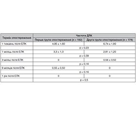
Актуальність. Дисемінований ламелярний кератит (ДЛК) є рідкісним, але тяжким післяопераційним ускладненням, що може виникнути після ексимерлазерної корекції короткозорості. За даними досліджень M. Moshirfar, K.M. Durnford, A.L. Lewis (2021), частота ДЛК після LASIK становить 4,3 % та 18,9 % — за даними Pranita Sahay, Rahul Kumar Bafna (2021). Частота ДЛК після FemtoLASIK (фемтосекундної ексимерлазерної корекції зору) становить від 0,5 до 37,5 % і більше за даними A. Leccisotti, S.V. Fields (2021). З огляду на зростаючу популярність технологій LASIK та FemtoLASIK, дослідження частоти і клінічних особливостей цього ускладнення є критично важливим для поліпшення результатів лікування і безпеки пацієнтів. Мета: дослідити особливості клінічного перебігу та частоту дисемінованого ламелярного кератиту після різних технологій ексимерлазерної корекції (ЕЛК) міопії. Матеріали та методи. За своїм дизайном це дослідження було проспективним, когортним, неінтервенційним. У дослідження було включено 180 пацієнтів (360 очей), які отримали корекцію короткозорості методом LASIK (182 ока) або FemtoLASIK (178 очей). Після операції проводився моніторинг, що включав візіометрію, рефрактометрію та біомікроскопію, для виявлення можливих ускладнень, зокрема ДЛК. Вивчали частоту та особливості клінічного перебігу після різних видів ЕЛК. Термін спостереження — 1 рік. Результати. При обстеженні через 1 тиждень після ЕЛК у пацієнтів першої групи спостереження частота ДЛК становила 4,95 %, у пацієнтів другої групи — 6,74 %. Через 1 місяць після ЕЛК було відмічено зменшення частоти ДЛК: у першій групі до 3,30 %, у другій групі — до 2,81 %. При обстеженні пацієнтів через 3 місяці після ЕЛК у першій групі спостереження проявів ДЛК не було, у другій групі спостереження його частота становила 0,56 %. При огляді через 6 місяців після ЕЛК в першій групі спостереження частота ДЛК становила 0,55 %, у другій групі клінічних проявів ДЛК не було. При огляді через 1 рік у пацієнтів першої та другої груп спостереження клінічних проявів ДЛК не було. Клінічний перебіг та прояви ДЛК після LASIK та FemtoLASIK не відрізнялись упродовж всього терміну спостереження. Також було встановлено, що 38,46 % пацієнтів мали в анамнезі SARS-CoV-2. Висновки. У результаті проведеного нами дослідження було встановлено, що частота ДЛК після LASIK була 6,3 %, а після FemtoLASIK — 6,6 %, при терміні спостереження 1 рік. Клінічний перебіг та прояви ДЛК після LASIK та FemtoLASIK не відрізнялись на всіх термінах спостереження. Було встановлено, що 38,46 % пацієнтів, яким була виконана ЕЛК за різними технологіями і у яких потім розвинувся ДЛК, мали в анамнезі SARS-CoV-2 (від 2 тижнів до 2 місяців). Подальшими перспективами нашого дослідження ми вважаємо вивчення нових місцевих (з боку органа зору) та загальних (з боку всього організму) патогенетичних чинників ДЛК після сучасних методів ексимерлазерної корекції аметропії.
Background. Diffuse lamellar keratitis is a rare yet severe postoperative complication that may arise following excimer laser correction of myopia. Research indicate that the incidence of this condition after LASIK ranges from 4.3 to 18.9 %, and after FemtoLASIK, it varies from 0.5 % to more than 37.5 %. Given the increasing popularity of LASIK and FemtoLASIK technologies, studying the frequency and clinical characteristics of this complication is crucial for enhancing treatment outcomes and patient safety. This study purposed to explore the clinical course and frequency of diffuse lamellar keratitis following different excimer laser technologies used for myopia correction. Materials and methods. We conducted a prospective, cohort, non-interventional study. It involved 180 patients (360 eyes) who underwent myopia correction using either LASIK (182 eyes) or FemtoLASIK (178 eyes). Postoperative monitoring included visual acuity, refraction, and biomicroscopy to identify potential complications, particularly diffuse lamellar keratitis. We examined the frequency and clinical course of the condition after each type of excimer laser correction. The observation period is 1 year. Results. Upon examination one week after excimer laser correction, the incidence of diffuse lamellar keratitis in the first group was 4.95 %, while in the second group it was 6.74 %. One month after surgery, there was a reduction in the frequency of diffuse lamellar keratitis: in the first group, it decreased to 3.30 %, and in the second group, to 2.81 %. By the three-month follow-up, the first group showed no manifestations of diffuse lamellar keratitis, whereas the second group had an incidence of 0.56 %. At the six-month follow-up, the incidence in the first group was 0.55 %, and there were no clinical manifestations of diffuse lamellar keratitis in the second group. At the one-year follow-up, neither group exhibited clinical signs of this disease. The clinical course and manifestations of diffuse lamellar keratitis did not differ between LASIK and FemtoLASIK throughout the study period. Additionally, it was found that 38.46 % of the patients had a history of SARS-CoV-2 infection. Conclusions. Our research revealed that the frequency of diffuse lamellar keratitis was 6.3 % after LASIK and 6.6 % after FemtoLASIK over a 1-year period. The clinical course and manifestations of the condition were similar for both LASIK and FemtoLASIK at all observation points. In was found that 38.46 % of patients who developed diffuse lamellar keratitis after excimer laser correction had a history of SARS-CoV-2 infection (from 2 weeks to 2 months). Future research should focus on investigating new local (ocular) and systemic (whole body) pathogenetic factors of diffuse lamellar keratitis following modern excimer laser methods for ametropia correction.
аномалії рефракції; ексимерлазерна корекція зору; ускладнення; дисемінований ламелярний кератит
refractive errors; excimer laser vision correction; complications; diffuse lamellar keratitis
Для ознакомления с полным содержанием статьи необходимо оформить подписку на журнал.
- Al-Haddad C., Hoyeck S., Torbey J., Houry R., Boustany R.N. Eye Tracking Abnormalities in School-Aged Children With Strabismus and With and Without Amblyopia. J Pediatr Ophthalmol Strabismus. 2019. 56(5): 297-304. DOI: 10.3928/01913913-20190726-01.
- Burton M.J., Loughnan B., Ervin M., et al. The Lancet Global Health Commission on Global Eye Health: vision beyond 2020. The Lancet Global Health. 2021. 9(4): e489-e551.
- Mitchell G.L., Rosner B., Richdale K. The development of a scoring algorithm for the Contact lens Assessment and Risk CARE Report. Optom Vis Sci. 2018. 95(Eabstract 180032).
- Rueff E.M., Wolfe J., Bailey M.D. A study of contact lens compliance in a nonclinical setting. Cont Lens Anterior Eye. 2019. 42(5): 557-561. DOI: 10.1016/j.clae.2019.03.001.
- Farooqui J.H., Acharya M., Kekan M. Current trends in surgical management of myopia. Community Eye Health. 2019. 32(105): S5-S6.
- Price M.O., Price D.A., Bucci F.A. Jr., Durrie D.S., Bond W.I., Price F.W. Jr. Three-Year Longitudinal Survey Comparing Visual Satisfaction with LASIK and Contact Lenses. Ophthalmology. 2016. 123(8): 1659-1666. DOI: 10.1016/j.ophtha.2016.04.003.
- Bowes Hamill M., Moshirfar M., Tarrant J., et al. AAO Refractive surgery BCSC 2019-2020. 2020. P. 130-147. DOI: 10.1016/S0140-6736(18)33209-4.
- Xia L.K., Yu J., Chai G.R., Wang D., Li Y. Comparison of the femtosecond laser and mechanical microkeratome for flap cutting in LASIK. Int J Ophthalmol. 2015. 8(4): 784-790. DOI: 10.3980/j.issn.2222-3959.2015.04.25.
- Mogilevskyy S.Yu., Zhovtoshtan M.Yu. Assessing the early and late impact of excimer laser correction for myopia on the development of dry eye syndrome. J Ophthalmol (Ukraine). 2022. 5: 23-29. DOI: 10.31288/oftalmolzh202252329.
- Pidro A., Biscevic A., Pjano M.A., Mravicic I., Bejdic N., Bohac M. Excimer Lasers in Refractive Surgery. Acta Inform Med. 2019. 27(4): 278-283. DOI: 10.5455/aim.2019.27.278-283.
- Kuryan J., Cheema A., Chuck R.S. Laser-assisted subepithelial keratectomy (LASEK) versus laser-assisted in-situ keratomileusis (LASIK) for correcting myopia. Cochrane Database Syst Rev. 2017. Issue 2: CD011080. DOI: 10.1002/14651858.CD011080.pub2.
- Kahuam-López N., Navas A., Castillo-Salgado C., Graue-Hernandez E.O., Jimenez-Corona A., Ibarra A. Laser-assisted in-situ keratomileusis (LASIK) with a mechanical microkeratome compared to LASIK with a femtosecond laser for LASIK in adults with myopia or myopic astigmatism. Cochrane Database Syst Rev. 2020. Issue 4: CD012946. DOI: 10.1002/14651858.CD012946.pub2.
- Spadea L., Giovannetti F. Main Complications of Photorefractive Keratectomy and their Management. Clin Ophthalmol. 2019. 13: 2305-2315. DOI: 10.2147/OPTH.S233125.
- Gaeckle H.C. Early clinical outcomes and comparison between trans-PRK and PRK, regarding refractive outcome, wound healing, pain intensity and visual recovery time in a real-world setup. BMC Ophthalmol. 2021. 21: 181. DOI: 10.1186/s12886-021-01941-3.
- Chang J.Y., Lin P.Y., Hsu C.C., Liu C.J. Comparison of clinical outcomes of LASIK, Trans-PRK, and SMILE for correction of myopia. J Chin Med Assoc. 2022. 85(2): 145-151. DOI: 10.1097/JCMA.0000000000000674.
- Wallerstein A., Kam J.W.K., Gauvin M., et al. Refractive, visual, and subjective quality of vision outcomes for very high myopia LASIK from −10.00 to −13.50 diopters. BMC Ophthalmol. 2020. 20: 234. DOI: 10.1186/s12886-020-01481-2.
- Saad A., Narr J., Frings A., et al. Surgical outcomes of laser in situ keratomileusis (LASIK) in patients with stable systemic disease. Int Ophthalmol. 2024. 44: 119. DOI: 10.1007/s10792-024-02956-7.
- Alió J.L., Toprak I., Alrabiah H. Intraoperative Complications of LASIK and SMILE. In: Albert D.M., Miller J.W., Azar D.T., Young L.H. (eds). Albert and Jakobiec's Principles and Practice of Ophthalmology. Springer, Cham. 2022. DOI: 10.1007/978-3-030-42634-7_228.
- Tamimi A., Sheikhzadeh F., Ezabadi S.G., Islampanah M., Parhiz P., Fathabadi A., et al. Post-LASIK dry eye disease: A comprehensive review of management and current treatment options. Front Med. 2023. 10: 1057685. DOI: 10.3389/fmed.2023.1057685.
- Ivarsen A., Hjortdal J. Complications and Management of SMILE. In: Linke S., Katz T. (eds). Complications in Corneal Laser Surgery. Springer, Cham. 2016. DOI: 10.1007/978-3-319-41496-6_10.
- Han T., Zhao L., Shen Y., Chen Z., Yang D., Zhang J., et al. Twelve-year global publications on small incision lenticule extraction: A bibliometric analysis. Front Med. 2022. 9: 990657.
- Moshirfar M., Durnford K.M., Lewis A.L., Miller C.M., West D.G., Sperry R.A., et al. Five-Year Incidence, Management, and Visual Outcomes of Diffuse Lamellar Keratitis after Femtose–cond-Assisted LASIK. J Clin Med. 2021. 10: 3067. DOI: 10.3390/jcm10143067.
- Diffuse lamellar keratitis after LASIK with low-energy femtosecond laser. J Cataract Refract Surg. 2021. 47(2): 233-237. DOI: 10.1097/j.jcrs.0000000000000413.
- Grassmeyer J.J., Goertz J.G., Baartman B.J. Diffuse Lamellar Keratitis in a Patient Undergoing Collagen Corneal Cross-Linking 18 Years After Laser In Situ Keratomileusis Surgery. Cornea. 2021. 40(7): 917-920. DOI: 10.1097/ICO.0000000000002653.
- Vera-Duarte G.R., Guerrero-Becerril J., Müller-Morales C.A., Ramirez-Miranda A., Navas A., Graue-Hernandez E.O. Delayed-onset pressure-induced interlamellar stromal keratitis (PISK) and interface epithelial ingrowth 10 years after laser-assisted in situ keratomileusis. Am J Ophthalmol Case Rep. 2023. 32: 101874. DOI: 10.1016/j.ajoc.2023.101874.
- Shetty A., Leal S., Mulpuri L., Tonk R. Late-onset Diffuse Lamellar Keratitis in the Context of Conjunctivitis Related to COVID-19: Case Report. J Refract Surg Case Rep. 2024. 4(1): e11-e14. DOI: 10.3928/jrscr-20240102-01.
- Karcenty M., Mazharian A., Courtin R., Panthier C., Guilbert E., Gatinel D. Management of epithelial ingrowth and diffuse lamellar keratitis caused by the interface penetration of an eyelash 12 years after laser in situ keratomileusis. J Fr Ophtalmol. 2022. 45(1): e43-e45. DOI: 10.1016/j.jfo.2021.02.005.
- Rosman M., Chua W.H., Tseng P.S.F., Wee T.L., Chan W.K. Diffuse lamellar keratitis after laser in situ keratomileusis associa–ted with surgical marker pens. J Cataract Refract Surg. 2008. DOI: 10.1016/j.jcrs.2008.02.014.
- Sahay P., Bafna R.K., Reddy J.C., Vajpayee R.B., Sharma N. Complications of laser-assisted in situ keratomileusis. Indian J Ophthalmol. 2021. 69(7): 1658-1669. DOI: 10.4103/ijo.IJO_1872_20.
- Yeoh C.B., Seier K.P., Francis J., Abramson D.H., Tan K.S., Tollinche L.E. Perioperative corneal injury: An unseen casualty of COVID-19. JOJ Ophthalmol. 2022. 9(2): 555757. DOI: 10.19080/jojo.2022.09.555757.
- Jiang L., Yang Y., Gandhewar J. Bilateral corneal endothelial fai–lure following COVID-19 pneumonia. BMJ Case Rep. 2021. 14: e242702.
- Wong L.R., Perlman S. Immune dysregulation and immunopathology induced by SARS-CoV-2 and related coronaviruses — are we our own worst enemy? Nat Rev Immunol. 2022. 22(1): 47-56. DOI: 10.1038/s41577-021-00656-2. Epub 2021 Nov 26. Erratum in: Nat Rev Immunol. 2022. 22(3): 200. DOI: 10.1038/s41577-021-00673-1.
- Akbari M., Dourandeesh M. Update on overview of ocular manifestations of COVID-19. Front Med (Lausanne). 2022. 9: 877023. DOI: 10.3389/fmed.2022.877023.
- Wong N.S.Q., Chang L., Lin M.T.Y., Lee I.X.Y., Tong L., Liu Y.C. Neuropathic Corneal Pain after Coronavirus Disease 2019 (COVID-19) Infection. Diseases. 2024. 12(2): 37. DOI: 10.3390/diseases12020037.
- Shetty A., Leal S., Mulpuri L., Tonk R. Neuropathic Corneal Pain after Coronavirus Disease 2019 (COVID-19) Infection. J Refract Surg Case Rep. 2024. 4(1): e11-e14. DOI: 10.3928/jrscr-20240102-01.
- Могилевский С.Ю., Павлюченко А.К. Причини неудач ексимер-лазерної корекції зору. Матеріали науково-практичної конференції офтальмологів з міжнародною участю «Філатовські читання», 28–29 травня 2009 р., Одеса. Одеса, 2009. С. 29.
- Могилевский С.Ю., Якубенко Е.Д., Павлюченко А.К. Особливості біохімічного статусу сльози у пацієнтів з міопією та міопічним астигматизмом і його вплив на частоту та характер ускладнень після ексимерлазерної корекції. Питання експериментальної та клінічної медицини: Збірник статей. Донецьк: ДонНМУ, 2010. Вип. 14. Т. 2. С. 208-213.
- Mogilevskyy S., Zhovtoshtan M., Bushuyeva O. Persistent dry eye syndrome after and late functional outcomes of excimer laser correction for myopia. J. Оphthalmol. (Ukraine) [Internet]. 2023 Feb. 28 [cited 2024 Sep. 3]. (1): 19-26. Available from: https://ua.ozhurnal.com/index.php/files/article/view/5.
- Wilson S.E., de Oliveira R.C. Pathophysiology and Treatment of Diffuse Lamellar Keratitis. J Refract Surg. 2020 Feb 1. 36(2): 124-130. doi: 10.3928/1081597X-20200114-01. PMID: 32032434.

